Activity 3.2: Tour d'Onion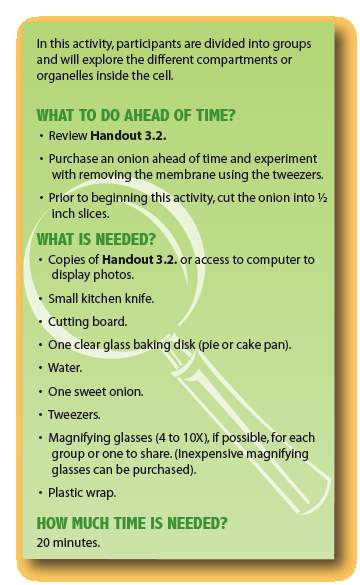
Directions
Remind participants that all fruits and vegetables, like humans, are made of cells (pass around an onion or several different types of onions to reinforce the diversity in onions that we learned about in Lesson 1, Activity 1.1).
- What do you think the different onions have that would make them look different from each other?
Invite them to plug their noses and go with you on a guided tour of the onion! Have participants form several teams, depending on how many materials you have. Distribute Handout 3.2 to each group. The leader distributes slices of the onion to each group. Starting at the center of a slice, have each group remove circular layers from the onion and place them on the bottom of a clear glass dish or plate with a thin layer of water. Ask each group to carefully remove membrane from the outside or inside of the circle using tweezers (Photograph 1) and place it on the water. Give each group a 6" piece of plastic wrap and ask them to touch the wrap to the membrane which will be floating on top of the water. The membrane will cling to the wrap and the group can lift the membrane out of the water. To prevent drying and to make it easier to view the membrane, fold the other side of the wrap over the exposed side of the membrane. If time is short the leader can "capture" the membrane ahead of time. Groups can then use their magnifying glasses to try to observe the outline of the cells in the membrane. Whether or not they can see the cells in the membrane, share Photograph 2. This is what they would see with a microscope that magnifies the image to 7 times its real size. Explain derivation of the word “cell” (see What's in a Word).
- Discuss why you think the "ancients" might have chosen the word, cell, to describe these structures?
- Can you describe what you might see inside the cell if you had a high powered microscope?
- Invite them to look at Photographs 3 through 7 in Handout 3.2. In these photographs different dyes are used to bind to certain structures in the cell. In Photograph 3 you see a slightly higher magnification of the onion cells in Photograph 2. In this image you can see dark green cell walls surrounding the outside of the stained green cells. These cells are 50-times larger than they actually are. If you enlarge the cells 200-times, shown in Photograph 4, you can see a smaller, bright green compartment inside the cell. That structure is considered to be the "control center" of the cell.
- Since this organelle is said to be the "control center", can you imagine what might be in this compartment?
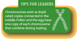
If you further magnify the brain center and apply a red stain, you can see some red, colored strands, the chromosomes (Photograph 5).
- Can you speculate as to what you think the structures are that are indicated by the arrows?
Using the dyes, other organelles besides the nucleus can be stained, like the sun energy capturer, the chloroplast (Photograph 6). You can also link specific pieces of DNA to a fluorescent dye, like the one in glow sticks. The piece of DNA scans the entire genome until it finds its match among the tens of thousands of genes in the cell and, when it is does, it sticks to the DNA and makes bright glowing spots along the chromosomal strands – so you can see exactly where that gene is, at 1000-times larger than their actual size (Photograph 7).
 To learn more about the structure of a plant cell and how it is different from an animal cell, click on the left image.
• Name two things that are different between a plant cell and an animal cell.